BACKGROUNDBrain edema is a severe complication of ischemic stroke that may result in secondary neurological deterioration and dying.
Glyburide is reported to forestall mind swelling in preclinical rodent fashions of ischemic stroke by way of inhibition of a non-selective channel composed of sulfonylurea receptor 1 and transient receptor potential cation channel subfamily M member 4. However, the relevance of this pathway to the event of cerebral edema in stroke sufferers is not recognized.
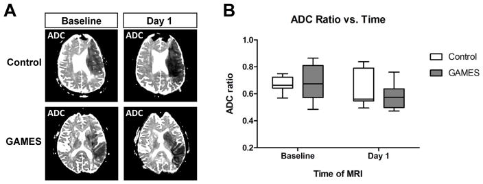
METHODSUsing a case-control design, we retrospectively assessed neuroimaging and blood markers of cytotoxic and vasogenic edema in topics who have been enrolled in the glyburide benefit in malignant edema and stroke-pilot (GAMES-Pilot) trial. We in contrast serial mind magnetic resonance photographs (MRIs) to a cohort with related massive quantity infarctions. We additionally in contrast matrix metalloproteinase-9 (MMP-9) plasma degree in massive hemispheric stroke.
RESULTSWe report that IV glyburide was associated with T2 fluid-attenuated inversion restoration sign depth ratio on mind MRI, diminished the lesional water diffusivity between days 1 and a pair of (pseudo-normalization), and lowered blood MMP-9 degree.CONCLUSIONSSeveral surrogate markers of vasogenic edema look like lowered in the setting of IV glyburide therapy in human stroke. Verification of those potential imaging and blood biomarkers is warranted in the context of a randomized, placebo-controlled trial.
Diffusion-weighted imaging exhibits cytotoxic and vasogenic edema in eclampsia.
In eclampsia, MR imaging exhibits reversible T2 hyperintensities in a parietal and occipital distribution. Findings on diffusion-weighted photographs counsel that these abnormalities are areas of vasogenic edema.
We describe the presence of each cytotoxic and vasogenic edema, as detected by diffusion-weighted imaging, in a lady with eclampsia. Follow-up MR imaging confirmed that the areas of cytotoxic edema progressed to cerebral infarction.
This case means that diffusion-weighted imaging permits the early detection of ischemic infarcts in sufferers with eclampsia.
Posterior reversible encephalopathy syndrome, half 2: controversies surrounding pathophysiology of vasogenic edema.
Posterior reversible encephalopathy syndrome (PRES) is a neurotoxic state accompanied by a singular mind imaging sample usually associated with numerous advanced scientific situations together with: preeclampsia/eclampsia, allogeneic bone marrow transplantation, strong organ transplantation, autoimmune ailments and excessive dose most cancers chemotherapy. The mechanism behind the growing vasogenic edema and CT or MR imaging look of PRES is not recognized.
Two theories have traditionally been proposed: 1) Severe hypertension results in failed auto-regulation, subsequent hyperperfusion, with endothelial harm/vasogenic edema and; 2) vasoconstriction and hypoperfusion results in mind ischemia and subsequent vasogenic edema.
The strengths/weaknesses of those hypotheses are reviewed in a translational vogue together with supporting proof and present obtainable imaging/scientific information associated to the situations that develop PRES.
While the hypertension/hyperperfusion principle has been hottest, the situations associated with PRES have the same immune problem current and develop the same state of T-cell/endothelial cell activation that could be the idea of leukocyte trafficking and systemic/cerebral vasoconstriction.
These systemic options alongside with present vascular and perfusion imaging options in PRES seem to render robust help for the older principle of vasoconstriction coupled with hypoperfusion because the mechanism.